- John Henry George Antrum, specialist registrar in plastic surgery1,
- Jennifer Eleanor Galloway, specialist registrar in emergency medicine2,
- Mohammad Umair Anwar, consultant burns and plastic surgeon1,
- Sophie Louise Hodson, foundation doctor3
1Pinderfields General Hospital Wakefield, Wakefield, UK
2Newcastle Upon Tyne Hospitals NHS Foundation Trust, Newcastle Upon Tyne, UK
3Hull Royal Infirmary Hull, Kingston upon Hull, UK
- Correspondence to: J H G Antrum henry.antrum{at}nhs.net
What you need to know
-
When calculating burn size, 1% of total body surface area (TBSA) is roughly equal to the size of the patient’s hand, including digits
-
Do not include areas of erythema without skin loss when calculating burn size
-
Small burns <2% TBSA in children and <3% TBSA in adults can be managed in local accident and emergency departments, minor injuries centres, or primary care practices if the patient is clinically stable and there are no complications or associated injuries
-
Cooling of the burn is effective up to 3 hours after the injury
-
Flamazine (silver sulphadiazine) cream is avoided except for infected burns as it is absorbed by the burnt skin and makes depth estimation difficult. It can also delay healing
Burn injuries are a common cause of presentation to hospitals and urgent care centres globally.1 About 137 000 patients attended accident and emergency (A&E) departments in England with a burn or a scald in 2019.2 Approximately 64 000 children seek medical attention for a burn from hospital or primary care centres in the UK each year.3 In the United States, 486 000 patients received medical treatment for burn wounds, which constituted approximately 2% of all emergency department attendances in 2017.4
Most burns can be managed in an outpatient setting with attentive wound care and regular changes of dressings. Inaccurate estimation of the burn size can result in inappropriate referrals to burns centres. Mismanagement of the wound increases the potential for prolonged pain,5 delayed healing, infection,6 and excessive scarring.7
This clinical update consolidates recent guidelines to help clinicians working in primary and urgent care facilities with assessment and management of small burn wounds. The management of burns from chemical and electrical injuries is not covered in this article.
What is a small burn?
There is no clear definition for a small or a large burn. Burns that are ≥10% of the total body surface area (TBSA) in children and ≥15% TBSA in adults require fluid resuscitation and expeditious transfer to a specialist burns service. Burns that are smaller than this can, in theory, be managed in A&E or primary care. In practice, however, burns greater than 2% TBSA in children and 3% TBSA in adults benefit from management and follow-up at a burns service. This is reflected in the UK guidelines for referral.8 For this article, we consider a small burn to be a cutaneous injury that is <2% TBSA in children and <3% TBSA in adults. However, even very small areas of cutaneous involvement can have severe consequences in the context of chemical or electrical injuries, infections, and smoke inhalation and need special attention.
What are the common types of burns?
Scalds and contact burns are the most common type of burn injury in both children and adults. Flame, flash (brief but intense exposure to heat), and chemical burns are more common in adults than children.9 Flame burns are proportionally much more common in developing countries.10
Contact burns—Commonly occur following contact with fire guards, radiators, electric irons, hair straighteners, and kitchen hobs that have not yet cooled. These are often superficial because the reflex withdrawal response limits contact time. Deep burns can occur in those with sensory deficits or episodes of unconsciousness. Deeper burns in vulnerable groups of people raise the possibility of non-accidental injury.
Flame burns—Clothes are protective but can cause deep burns if ignited and not removed quickly. Flame burns with petrol are deeper than those without and are at a higher risk for inhalation injury when in an enclosed space. In an explosion, consider blunt force traumatic injuries in addition to the burn.
Scalds—Caused by hot liquids. Thicker liquids (such as soup) retain heat for longer, causing deeper burns. Ask how recently the liquid was prepared to estimate heat at the point of contact. Again, clothes are protective if removed quickly but can deepen the burn with prolonged contact. Elderly patients can present with deep scalds to the thighs, genitals, and lower abdomen if they have been unable to remove their clothes themselves.
Chemicals—Chemical burns can result from substances used in industry, agriculture, and domestic cleaning.
Electrical injuries—These are divided into low voltage (<1000 V), high voltage (>1000 V), and lightning strikes. Domestic appliances are low voltage (230 V in Europe and 120 V in North America) and have potential to cause significant local cutaneous (entry and exit) wounds and cardiac arrest but less deep tissue damage. High voltage injuries can cause substantial cutaneous and soft tissue damage affecting multiple organ systems. Wet skin can convert a small injury into a serious one.
What are the key features of an initial assessment?
First distinguish minor injuries from potentially life or limb threatening injuries. People with major burns or traumatic injuries are usually identified early and brought directly to A&E by ambulance, though a patient may be unwell with small cutaneous burns, especially with infections, inhalational injuries and electrical injuries.
A focused burns history will help determine the severity of burn, identify any associated injuries, and determine the need for referral.
Ask how the burn was sustained as burns can occur in the context of other injuries or illnesses, such as collapse (due to a neurological or cardiac condition), or self harm. Detailing the events around the burn will determine if additional investigations or specialist support are required.
Ask when and where the burn was sustained. Patients usually present immediately after sustaining a burn, especially if it is relatively large or painful. Some try to manage the burn themselves initially, with ointments from a chemist or anecdotal home remedies (such as toothpaste or yogurt) and only attend hospital when the burn has changed or become unmanageable. Burns that do not heal within three weeks are at high risk of prominent scarring.711 If the burn happened outdoors or in an unclean environment, then the wound may be contaminated and at risk of infection. Flame burns that occur in confined spaces raise the likelihood of an associated inhalational injury.
Elicit the patient’s vaccination history along with occupation and hand dominance for upper limb burns. Ask about other medical conditions. Patients who struggle with independence will need special arrangements for follow-up.
What are the special considerations in children presenting with burns?
Safeguarding the child is a priority in all paediatric consultations. The possibility of neglect or abuse should always be considered in children and vulnerable populations. Most injuries will be accidental, but any concerning features must be discussed with a paediatrician (box 1). Be mindful that even considerate parents struggle with childcare for various social reasons, and there may be an opportunity to educate the parent(s) or provide additional support in the form of a social worker.
“Red flags” for non-accidental injury
-
Absent, inconsistent, or unfeasible explanation for the injury
-
History inconsistent with the child’s level of mobility or examination findings
-
Injury to body areas unlikely to be exposed to hot objects
-
Burn wound in the shape of an object
-
Delayed presentation (but may also be due to attempts at first aid)
-
Lack of parental concern
-
Failure to engage with healthcare professionals
-
Associated injuries unrelated to the burn (including possible signs of restraint)
-
A history of non-engagement, social service involvement, or “red flags”
How do I assess a burn wound?
Burn size
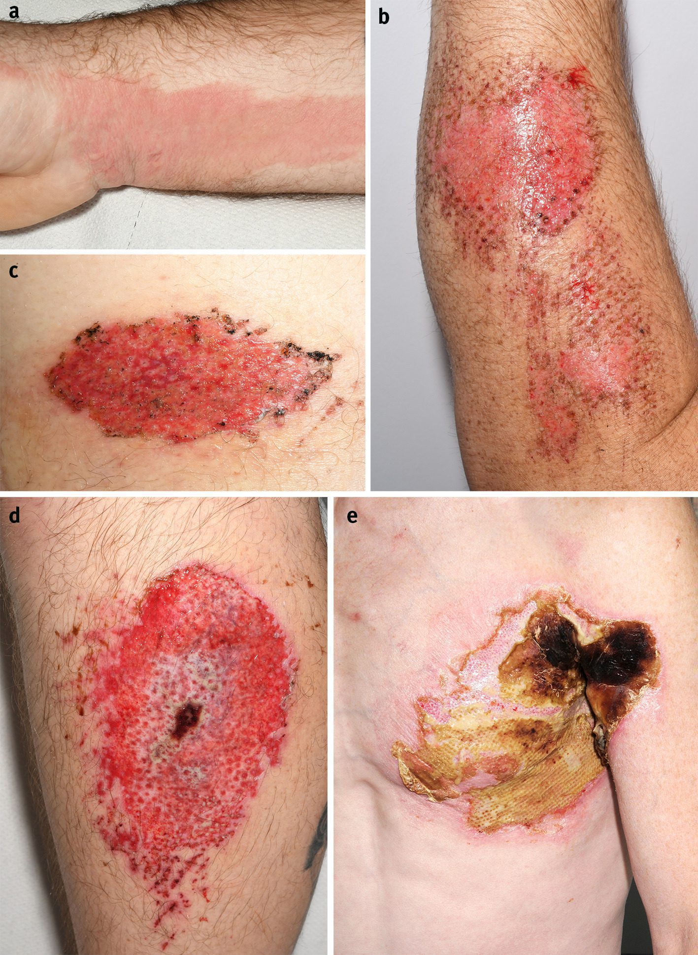
Burns are calculated as a percentage of the patient’s total body surface area. Pure erythema (superficial burns without blistering) is not included in the calculation1213; this results in overestimation of the burn size. There are various diagrams to assist with burn size estimation, the most well-known being “Wallace’s Rule of 9’s” and the “Lund & Browder Chart.”13 The latter is considered more accurate in children as it accounts for the different body proportions. Smartphone apps can also assist with burn size estimation, such as the “Mersey Burns App,” which is endorsed by the British Association of Plastic and Reconstructive Surgeons (BAPRAS).1415 As a general rule, 1% is equal to the size of the patient’s hand (both the palm and fingers). Areas smaller than this can be described in conventional units of size.
Burn depth
The terminology used for burn depth varies internationally. The British Burn Association (BBA) describes burn depth according to the skin anatomy,16 whereas the American Burn Association (ABA) classifies depth in degrees17 (table 1).
Burn depth diagnosis and classification
Burn site
Burns to certain body regions can be particularly difficult to manage, namely the hands, feet, face, perineum, and genitalia. Facial burns can lead to significant swelling and may be accompanied by ocular or inhalational injuries. Burns to the hands can cause functional impairment, particularly with subsequent scar contractures, and their management often involves regular physiotherapy. Burns to the feet are also prone to swelling and may require a period of bed rest and elevation.18 Genital burns have potential to restrict micturition and may need catheterisation. Both perineal and genital burns are prone to contamination and require regular dressing changes.
A circumferential burn is present when the entire circumference of a limb (or the trunk, neck, or digits) is burned. The inelastic burned skin has potential to constrict the underlying tissues, particularly if tissue oedema progresses, leading to tissue necrosis. All circumferential burns should be discussed with a burns service urgently.
Clinical photographs of the burns (with the patient’s consent) are the most accurate way of documenting the burn and monitoring healing. They can also be sent to burns services for review if required.
When should I refer?
Consider referral to specialist burns services for patients having large or complex burn wounds, or associated complications (see box 2). The nearest burns service is usually the first point of contact for referrals, whatever level this may be (box 3). The burn wound can be covered with cling film during transfer.
Criteria for referral to a specialist burns service82122
-
Infected burns
-
An unwell child with a burn
-
Burns >2% of total body surface area in children or >3% in adults (remember not to include simple erythema)
-
Circumferential burns
-
Full thickness burns
-
Burns involving the face, hands, genitals, or perineum
-
Chemical, electrical, and friction burns along with cold injuries
-
Burns with concerns about non-accidental injury or neglect
-
Burns in patients with complex medical or social issues that could complicate treatment or recovery
-
Burns that are ≥2 weeks old and have not healed
Levels of burns services (UK)8
-
Burn facility
-
Part of a plastic surgery service
-
Inpatient care equivalent to a standard plastic surgical ward
-
Acute care for non-complex burns
-
-
Burns unit
-
Burns centre
Many services now use online referrals19 whereby clinical details and photographs can be sent directly from a computer or smartphone, or with live video assessment. This can reduce travel requirements for patients, especially in areas far from specialist centres.20
What are the first aid measures for burns?
Cool the burn with cool running water for 20 minutes. Cooling the burn is effective up to three hours after the injury.23 If the patient presents within that time, further cooling in the department will help to prevent progression of the burn. Cooling eases pain, reduces the inflammatory reaction, and prevents progression of the burn.24 Ice or iced water should not be used as this causes vasoconstriction and can deepen the burn along with increasing the risk of hypothermia.
Remove rings and jewellery near the burn25 as subsequent swelling risks vascular compromise of the digits.
How do I manage the burn wound?
No specific treatment is usually required for epidermal (first degree) burns. Moisturising cream can be applied. Ask the patient to return if blistering occurs later or the wound is very painful. Antibiotic ointments, hydrogels, petroleum jelly, or soft paraffin can be used for superficial facial burns.
For deeper wounds, provide suitable analgesia before cleaning. Remove wet or burnt clothing to access the burn and prevent hypothermia. Remove any debris from the area. Expose and clean one site at a time to reduce heat loss. Gently clean the burn with warm water and dilute aqueous chlorhexidine (0.1% or 0.2%)2627 as this has broad antimicrobial coverage. Soap with warm water or saline is an appropriate alternative if chlorhexidine is not available.
Remove any loose skin and blisters that have already ruptured.28 The removal of blisters facilitates accurate assessment of the burn size and depth, reduces the potential for infection, and increases the efficacy of topical ointments and antimicrobials. Most blisters will lift with cleaning. Generally, large or tense blisters should be removed in sterile conditions with scissors and forceps. Small (<6 mm), non-tense blisters can be left alone.
What dressings are used for burn wounds?
Dressings are used to protect the wound from further trauma and maintain an optimal environment for healing. Numerous dressings with different properties have been used to treat burn wounds, though the evidence for each is of low quality, as identified in various systematic reviews.29303132Box 4 lists the common types of dressings with branded examples available in the UK.
Common types of burn dressings and ointments with named examples
-
Antimicrobials*—Activon (honey), bacitracin, Flamazine (silver sulfadiazine), Flammacerium (silver sulfadiazine + cerium nitate)
-
Hydrocolloids—These absorb exudate to form a moist gel which encourages granulation. They do not adhere to the burn wound itself. A semi-occlusive film maintains moisture while providing a barrier to microbial colonisation. Aquacel†, Comfeel, Duoderm, UrgoClean†
-
Hydrogels—These have a similar composition to hydrocolloids with a high water content34 and rehydrate dry slough or eschar. Many formulations are amorphous and can conform to any wound bed. Prontosan*
-
Alginate dressings—These are derived from brown seaweed and are highly absorbent, also forming a gel when in contact with wound exudate. They tend to dry and adhere to the wound, which can make them unsuitable for burn wound management. Algivon* (honey), Flaminal Hydro*, Flaminal Forte*
-
Low adherence dressings—These consist of a fabric mesh or “tulle” (predominantly cotton or polyester) impregnated with a lubricant such as paraffin or triglycerides. They are suitable for wounds with low amounts of exudate, though care must be taken to prevent the mesh from drying and sticking to the wound bed during epithelialisation. They may need to be changed more frequently than other types of dressings. Acticoat*, Atrauman†, Bactigras*, Jelonet
-
Soft polymer dressings—Often a silicon polymer which gently adheres to the wound and is easily removed. Adaptic Touch, Mepilex†, Mepitel, Urgotul†
A primary, non-adherent layer should be in contact with the burn, and a secondary, absorptive layer on top to collect exudate. Some composite dressings contain both layers. Antimicrobial primary dressings are preferred33 as they reduce the risk of infection. Silver is often incorporated into a dressing because of its broad antimicrobial cover. Hydrocolloids, hydrogels, and alginates promote autolytic debridement of wound slough.
Silver sulfadiazine (SSD) ointment contains the sulfonamide broad-spectrum antibiotic and has been widely used in the treatment for burns. Its application is still advocated by the American Burn Association.27 Silver sulfadiazine forms a ‘pseudoeschar’ when in contact with a burn—a khaki staining of the wound itself which makes subsequent assessment difficult. The available reviews29303132 generally favour silver based dressings compared with silver sulfadiazine with regard to infection rates, pain, wound healing time, and frequency of dressings changes.
For clean dermal wounds, use a soft polymer or low adherence dressing (ideally with antimicrobial properties) depending on availability and patient preferences. Absorbent gauze should be added for moderate and highly exudative wounds. For wounds with small amounts of exudate, a composite dressing may be used. For infected or sloughy wounds, use antimicrobial dressings after consultation with a burns service. Honey and hydrocolloids aid debridement. Alginates can be used for highly exudative wounds.
What additional measures are advised?
Tetanus prophylaxis35—Burns that are heavily contaminated with environmental debris and those with extensive devitalised tissue are at high risk of tetanus infection.36 Consider whether a patient requires vaccination or immunoglobin as per national or regional tetanus guidelines.
Antibiotics—The routine use of prophylactic systemic antibiotics is not recommended for small burns in the acute setting.37 Some units advocate their use in children under 5 years old to prevent toxic shock syndrome, though this use is anecdotal.
What are the possible acute complications?
Infection
Infection is a leading cause of morbidity and mortality in patients with burns. Infection can deepen the burn wound and, in severe cases, cause systemic sepsis. Infection in the small burn wound presents with increased pain, localised cellulitis, warmth, induration, increased exudate (often malodourous), discoloration of the dressing, and a change in the appearance of the burn itself.37
Consult the nearest burns service for management or referral of patients with infected burn wounds. Mild localised infections can be managed with oral antibiotics in an outpatient setting, but more severe infections may require intravenous antibiotics and surgical debridement.
Toxic shock syndrome
Toxic shock syndrome (TSS) is an uncommon but severe systemic illness with a high mortality if left untreated. It is caused by exotoxins from colonising bacteria (most commonly Staphylococcus aureus) which provoke an overwhelming immune response in patients with impaired immunity.38 In the context of a burn, children under 4 years old are at greatest risk as they have not yet developed antibodies to the exotoxins, and the burned skin loses its protective barrier against the colonising bacteria. The typical presentation is a young child with sudden clinical deterioration one or two days after a burn injury. Clinical findings may include pyrexia (≥39°C), rash, lethargy, irritability, diarrhoea or vomiting, and lymphopenia.39 The burn wound itself may be clean and unremarkable. It is important to immediately consult both a paediatrician and a burns service for any unwell or pyrexial child who presents after a burn injury.
How do I monitor the burn wound?
For burns that do not require management at a burns service, follow-up can take place in primary care. Some A&E units have their own follow-up clinics. Arrange an initial review within 2-3 days to review depth progression, infection, and exudate. All burn wounds develop a paler appearance at day 2-3 due to tissue fluid and biofilm formation, which may be mistaken for burn wound progression or infection. Arranging the first dressings change at this point enables review of these changes.
Subsequent dressing changes can occur every 3-7 days.40 Low adherence or “tulle” dressings may need to be changed every 3-4 days to prevent wound adherence. Dressings without antimicrobial properties are also changed earlier than antimicrobial dressings. Superficial burns should heal within two weeks. If there are concerns about infection, deepening of the burn, or delayed healing, discuss this with the nearest burns service.
A patient’s perspective
After a night shift I fell asleep with a hot water bottle by my feet. I awoke with a strangely warm sensation on my ankle and removed the covers to see a blister (~20 mm diameter) on my ankle. I panicked and realised I had fallen asleep with my foot on the hot water bottle. I ran my foot under cold water. The duration of contact had been over five hours, so I was aware this may have little effect. I cleaned the area, not disturbing the blister and placed a burns plaster and dressing on my ankle. I treated my ankle in this manner for over a week, continuing to work all of my regular hospital shifts, before it was apparent that it had become infected. My ankle was red and swollen, and it was painful to walk.
I went to A&E, where the wound was debrided, and I was given antibiotics for the infection. Two weeks later, the burn was still not healing. I was referred to the plastics and trauma clinic. Because of the size and depth of the burn, I was advised to have a skin graft. I underwent the procedure, and I am pleased with the outcome.
In hindsight, I should have come to hospital immediately after waking and cooling my foot. This might have helped the wound to heal and could have prevented infection, delayed healing, and the need for a skin graft.
How patients were involved in the creation of this article
SLH is a patient co-author, who provided her own perspective of having a burn wound managed by both A&E and plastic surgery services. She supported the other authors by reviewing the initial draft of the article and checking that the considerations of treatment from a patient’s perspective had been addressed.
Education into practice
-
How will you evaluate a patient presenting with a burn wound?
-
When will you refer patients to a specialist burns service? How will you arrange referral with your nearest burns service?
-
What dressings do you commonly use at your practice for management of burns? How do you discuss these with your patient?
Footnotes
-
Contributors: JHGA and MUA conceived of the article. JHGA wrote the first draft and JEG edited this with additions from an emergency medicine perspective. SLH contributed the patient perspective. All authors approved the final version. JHGA is the guarantor.
-
Competing interests: We have read and understood BMJ policy on declaration of interests and have no relevant interests to declare.
-
Patient consent: Patient consent obtained.
-
Provenance and peer review: Commissioned; externally peer reviewed.
References
- ↵
- ↵
- ↵
- ↵
- ↵
- ↵
- ↵
- ↵
- ↵
International Burn Injury Database (iBID). Burn dashboard data for England & Wales 2019.
- ↵
- ↵
- ↵
Education Committee of the Australian and New Zealand Burn Association Limited. Burn wound assessment. In: Emergency Management of Severe Burns (EMSB) Course manual, 16th edition. 2016: 43.
- ↵
- ↵
- ↵
- ↵
Education Committee of the Australian and New Zealand Burn Association Limited. Burn wound assessment. In: Emergency Management of Severe Burns (EMSB) Course Manual, 16th edition. 2016: 42-44.
- ↵
- ↵
- ↵
- ↵
- ↵
- ↵
European Burns Association. Burn care provision recommendations for European minimum level of provisional burn care. In: European Practice Guidelines for Burn Care. 2017: 19-20.
- ↵
Education Committee of the Australian and New Zealand Burn Association Limited. Management of the burn wound. In: Emergency Management of Severe Burns (EMSB) Course Manual, 16th edition. 2016: 51.
- ↵
- ↵
- ↵
Education Committee of the Australian and New Zealand Burn Association Limited. The outpatient management of the minor burn. In: Emergency Management of Severe Burns (EMSB) Course Manual, 16th edition. 2016: 98.
- ↵
- ↵
- ↵
- ↵
- ↵
- ↵
- ↵
- ↵
- ↵
- ↵
- ↵
- ↵
- ↵
- ↵
Education Committee of the Australian and New Zealand Burn Association Limited. The outpatient management of the minor burn. In: Emergency Management of Severe Burns (EMSB) Course Manual, 16th edition. 2016: 100.


